Gum Tissue Around Dental Implants in Japan: How Much Matters
Reviewed by Dr. Ryosuke Murai, DDS, MSD — Prosthodontist, trained at Indiana University School of Dentistry (U.S.). Last updated August 2026.
A patient arrives with an implant placed elsewhere three years ago. The implant is solid, the crown is intact, the X-ray looks acceptable — and yet the area is sore every time she brushes it, and the metal collar has started to show. Nothing failed. What she is missing is a band of firm, attached gum around the implant. The amount and quality of gum tissue around a dental implant is one of the least discussed variables in implant planning, and one of the few that is far cheaper to address before the crown goes on than after. This guide is for self-pay patients who want to understand what the tissue does, how much of it the evidence supports, and when grafting is genuinely worth it. It sits alongside our wider explanation of how implant cases are planned by a U.S.-trained prosthodontist.
Why Gum Around an Implant Is Not Gum as You Know It
Around a natural tooth, fibers from the periodontal ligament fan out and insert into the root surface, anchoring the gum and giving it a mechanical seal. An implant has no periodontal ligament. The tissue around it attaches in a weaker, more parallel arrangement, with a poorer blood supply. That is the whole reason this topic exists: the same amount of tissue does less work around an implant than it does around a tooth.
Two properties matter, and they are frequently confused. Keratinized tissue is the tough, pale, stippled band that resists the abrasion of a toothbrush — as opposed to the darker, looser lining mucosa deeper in the cheek. Mucosal thickness is a separate measure: how bulky the tissue is from outside to inside, which governs whether grey metal shows through and how the margin looks. A site can have a wide keratinized band that is paper-thin, or thick tissue with almost no keratinized band. They are addressed by different procedures.
How Much Is Enough
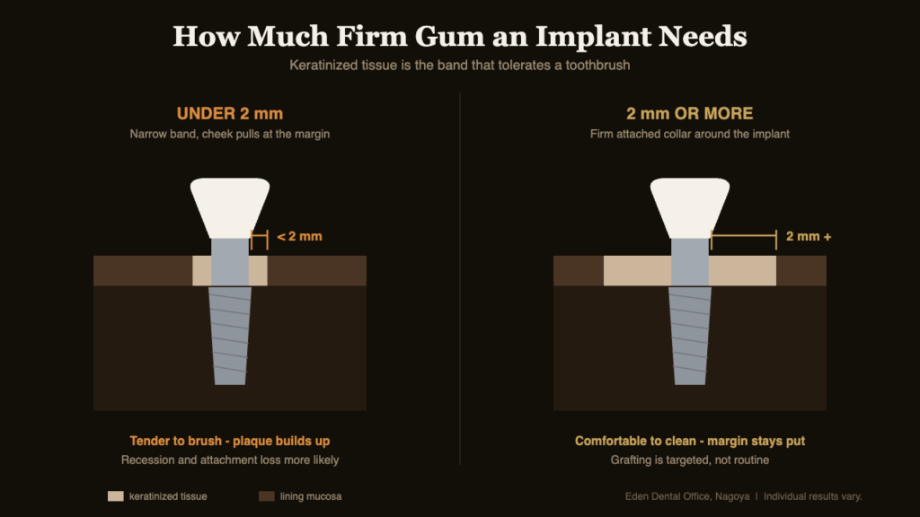
The threshold used in most of the literature and in our own planning is 2 mm of keratinized tissue, of which at least 1 mm is firmly attached rather than mobile. Below that, brushing the area tends to be uncomfortable, the tissue is pulled by the cheek and lip during function, and day-to-day plaque control quietly degrades — which is the mechanism that matters, rather than the width itself.
| Situation | What tends to follow | Usual response |
|---|---|---|
| ≥2 mm keratinized, thick tissue | Comfortable brushing, stable margin | No intervention |
| ≥2 mm keratinized, thin tissue | Metal or grey shadow may show through | Thickening graft if the site is visible |
| <2 mm keratinized, back teeth | Tender brushing, plaque accumulation | Widening graft, usually at uncovering |
| <2 mm keratinized, front teeth | Recession risk in the smile line | Graft before the final crown is made |
| Lower back jaw, muscle pull at the site | Tissue lifted away from the implant | Widening graft; results are least predictable here |
What the Evidence Supports — and What It Does Not
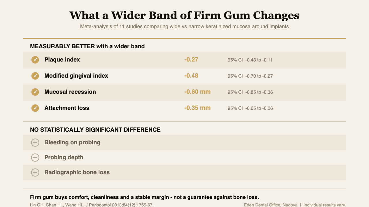
A systematic review of eleven studies compared implants with wide versus narrow keratinized mucosa. Implants with a wide band showed significantly better plaque index (weighted mean difference -0.27, 95% CI -0.43 to -0.11), modified gingival index (-0.48, 95% CI -0.70 to -0.27), mucosal recession (-0.60 mm, 95% CI -0.85 to -0.36) and attachment loss (-0.35 mm, 95% CI -0.65 to -0.06). Just as importantly, bleeding on probing, probing depth and radiographic bone loss showed no statistically significant difference (J Periodontol 2013).
Read that honestly. Keratinized tissue buys comfort, cleanliness and a more stable gum margin. It has not been shown to be the thing that decides whether you lose bone. Anyone selling a graft as insurance against implant failure is going further than the data allows.
Grafting itself has been examined the same way. A systematic review screening 2,823 studies found that procedures to gain keratinized tissue produced significantly better gingival index scores than leaving the site alone (weighted mean difference 0.863, 95% CI 0.658 to 1.067, p < .001), and that an apically positioned flap combined with an autogenous graft gave better final marginal bone levels than the comparison treatments (-0.175 mm, 95% CI -0.313 to -0.037, p = .013). Procedures aimed only at increasing mucosal thickness did not significantly improve bleeding indices over time (Clin Oral Implants Res 2018). Grafting works, in other words, but for specific and fairly modest endpoints.
When We Graft, and When We Leave It Alone
At Eden the tissue is assessed twice: once at planning, when we measure the keratinized band and the thickness on the intraoral scan and probe the site, and again at uncovering, when the healing abutment is placed and the tissue can be moved. Most grafting, when it is needed, happens at that second moment — the site is already being opened, so the extra procedure adds one appointment rather than a separate surgical episode. Where thickness rather than width is the problem, a connective tissue graft taken from the palate is placed under the tissue at the same visit.
We do not graft as a default. A molar site with a firm 2 mm band and no muscle pull is left alone, because a graft there has a donor site, swelling and cost attached to it for a benefit the patient will never notice. The sites where we do recommend it are the ones the table above describes: visible front teeth, thin tissue over a metal component, and lower back segments where the cheek muscle actively lifts the tissue away. The decision is easier and cheaper before the crown is made — once the final crown and its gum contours have been shaped and recorded, changing the tissue means remaking the restoration – a cost that depends heavily on whether that crown was screw-retained or cemented.
This is also why we take the soft tissue seriously at the very first planning stage rather than treating it as a finishing detail. The position of the fixture determines how much tissue is available around it, so the assessment belongs with the CBCT and scan-based plan rather than after it. For patients traveling in for treatment, knowing at the planning stage whether a graft is likely also changes the shape of the trip, which we cover in our guide to the implant procedure and recovery.
Frequently Asked Questions
Do all dental implants need a gum graft?
No. Most sites with a firm band of at least 2 mm and no muscle pull need nothing. Grafting is targeted at thin, narrow or visible sites.
Does a gum graft stop an implant from failing?
The evidence does not support that claim. Wider keratinized tissue improves plaque control, comfort and recession, but has not been shown to change radiographic bone loss.
Where does the graft tissue come from?
Usually your own palate. Collagen matrix substitutes exist and avoid a donor site, though autogenous tissue performed better for marginal bone levels in the reviewed studies.
When should grafting be done around an implant?
Usually at uncovering, when the healing abutment is placed. Doing it then adds one appointment instead of a separate surgery, and precedes crown design.
Can gum be added years after the crown is fitted?
Sometimes, but the crown often has to be removed or remade to gain access and to match the new margin, so the cost is considerably higher.
What This Costs
Soft-tissue grafting is quoted per site and depends on whether the aim is width or thickness, whether tissue is taken from your palate or a substitute material is used, and whether it can be combined with an appointment you were having anyway. Because it is not a fee we publish as a fixed figure, our fee page explains how self-pay pricing is structured, and every plan is issued as a written estimate in English, itemized by stage, before treatment begins. Each case starts with a comprehensive examination and records (check-up package ¥40,000). The practical point on cost is timing rather than technique: grafting folded into the uncovering appointment is markedly cheaper than grafting arranged after a final crown exists, and cheaper again than the long-run maintenance of a site that is uncomfortable to clean.
Risks and limitations. Soft-tissue grafting involves a surgical procedure with swelling, bruising and post-operative discomfort, and where tissue is taken from the palate there is a second wound that is usually the more uncomfortable of the two for the first week. Grafts can shrink over the months that follow, and the amount of gain is not fully predictable; the lower back jaw is the least predictable region. Results also depend on smoking status, oral hygiene and general health. The published benefits concern plaque control, gingival inflammation scores and recession — not implant survival. Individual results vary. Whether your site needs grafting can only be judged from an examination with records and probing measurements.
If you are planning implant treatment from elsewhere in Japan or from overseas and want the soft-tissue plan explained before you travel, our implant treatment page sets out how cases are assessed here, and our English-speaking team answers questions directly on WhatsApp.
More on this topic: Implant Guides — every article we have published in this cluster, grouped by stage.