
CBCT Scans Before Dental Implants in Japan: What We See
Reviewed by Dr. Ryosuke Murai, DDS, MSD — Prosthodontist, trained at Indiana University School of Dentistry (U.S.). Last updated August 2026.
A CBCT scan is a three-dimensional X-ray of the jaw, taken in about ten to twenty seconds while you sit upright, and in Japan it is the diagnostic step that determines whether a dental implant can be placed, exactly where it can sit, and whether bone grafting is needed first. It is a different examination from the flat panoramic image most patients have already had. This guide explains what a CBCT scan for dental implants in Japan measures, how three-dimensional information changes a treatment plan, what the radiation exposure means in practical terms, and what to do if you already hold a scan from a clinic in Tokyo, Osaka or abroad. It is written for self-pay patients who want to understand a recommendation before agreeing to it.
The X-ray you already have may not settle the question
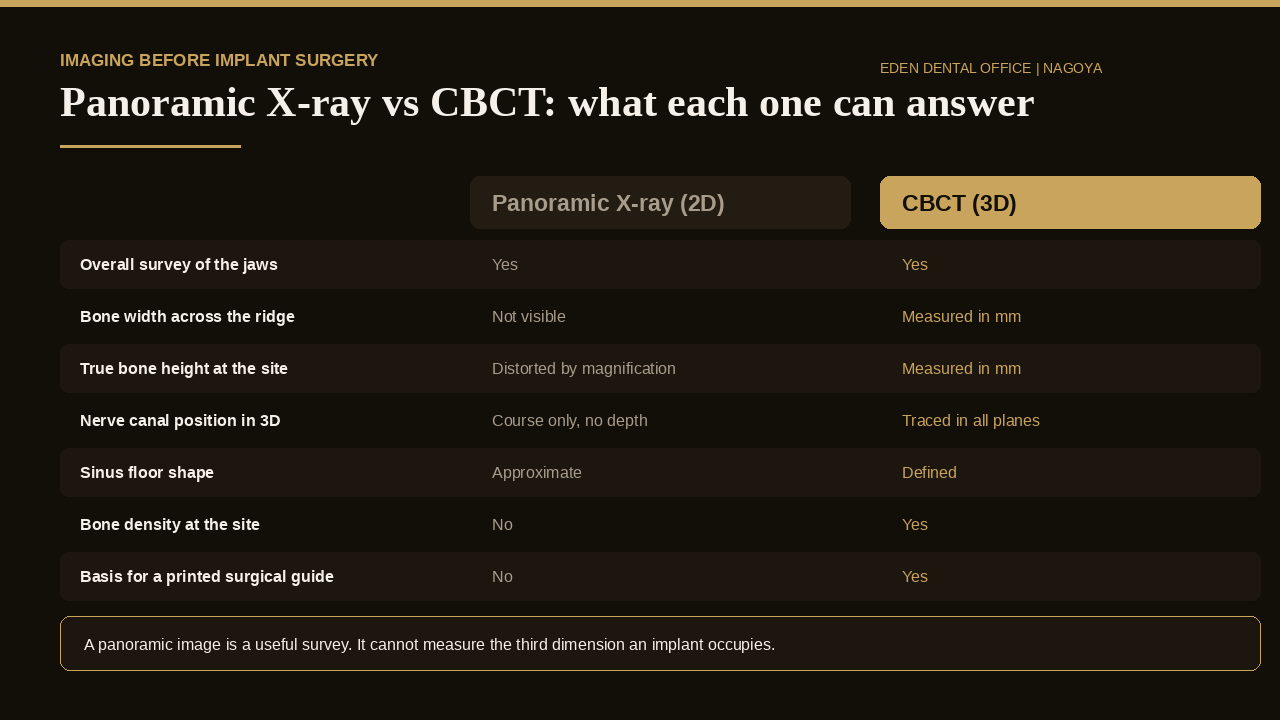
Most patients arrive holding a panoramic radiograph — a useful survey image showing every tooth, the outline of both jaws and obvious pathology in one frame. What it cannot do is measure the dimension an implant occupies. A panoramic image is a flattened projection, magnified unevenly across its width, so a ridge that looks generous on film may be a knife-edge of bone in cross-section, and a nerve canal that appears to run safely below the site may sit toward the cheek or the tongue side of it.
This is not a theoretical distinction. In a study published in Clinical Oral Investigations, clinicians planned the same cases twice, once from panoramic images and once from CBCT, and their decisions changed: they tended to choose larger implant dimensions from the panoramic view and smaller ones once they could see the site in three dimensions, and less experienced clinicians who had planned a bone graft from the flat image often withdrew it after viewing the scan. The imaging did not confirm the plan. It rewrote it — in both directions.
Five measurements that decide where an implant goes
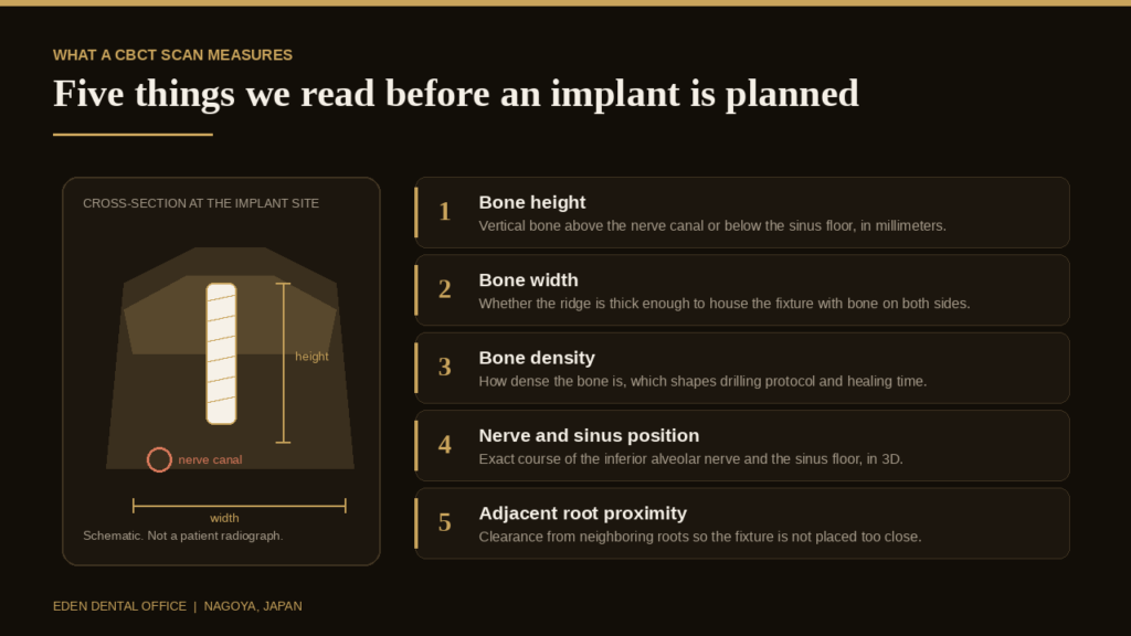
Bone height is the vertical distance above the inferior alveolar nerve in the lower jaw, or below the sinus floor in the upper, measured in millimeters at the exact planned site rather than averaged across the region. Bone width is the thickness of the ridge in cross-section, which decides whether a fixture can be housed with bone remaining on both the cheek and tongue surfaces. Width is the measurement panoramic imaging does not contain, and it is the most common reason a case that looked straightforward becomes a discussion about grafting and its separate fee.
Bone density influences the drilling sequence, the initial stability we can expect on the day, and how long we wait before loading. Nerve and sinus position are traced in all three planes rather than estimated, which allows a safety margin to be defined numerically instead of by eye. Adjacent root proximity tells us whether neighboring roots leave enough clearance, or whether the fixture must be angled or moved. The same measurements drive candidacy in full-arch cases, where bone volume is usually less decisive than patients expect.
What a finding on the scan actually changes
The value of the scan is not the image but the decision that follows it. The table below sets out the findings we most often act on.
| What the scan shows | What changes in the plan |
|---|---|
| Adequate height and width at the site | Straightforward placement; guide designed to the planned position |
| Thin ridge, adequate height | Narrower fixture, ridge expansion, or guided bone regeneration alongside placement |
| Reduced height below the sinus | Crestal or lateral sinus floor elevation, quoted as its own line item |
| Nerve canal closer than expected | Shorter fixture, repositioned site, or a different tooth-replacement option |
| Low bone density | Modified drilling protocol and a longer healing interval before the crown |
| Infection or a retained root fragment | Site treated and allowed to heal before any implant is scheduled |
Several of these outcomes make treatment simpler and cheaper than the initial assumption, which is worth stating plainly: a scan is not a device for adding procedures to a quote. It is how the sequence in our guide to the implant procedure, timeline and recovery is fixed before anyone picks up an instrument.
Radiation, in plain terms
This is the question patients ask most often, and it deserves a direct answer rather than reassurance. A dental CBCT is a small-volume scan of the jaws, not a medical CT of the head, so the exposure is correspondingly limited — but it is not a single fixed figure. A systematic review in the International Journal of Oral & Maxillofacial Implants covering guidelines, indications and dose risk in implant dentistry found that effective doses across CBCT devices span roughly a hundredfold from the lowest-dose unit to the highest, and that meaningful reduction is achieved by adjusting exposure settings and restricting the field of view to the region actually under investigation.
Two practical consequences follow. A scan should be justified — taken because a specific question needs answering, not added routinely to every appointment. And the field of view matters: a scan limited to one quadrant delivers considerably less exposure than a full-jaw volume, so it is reasonable to ask which is being used and why. Patients who are pregnant, or think they may be, should say so before any radiographic examination.
From the scan to a printed surgical guide
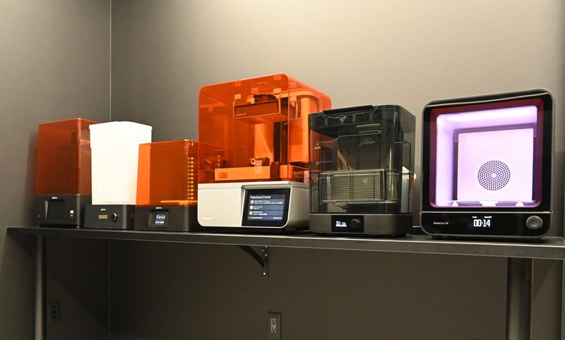
At Eden, a CBCT scan is never read in isolation. The DICOM data is merged with an intraoral scan and with the position the final tooth needs to occupy, so the implant is planned backward from the restoration rather than forward from whatever bone happens to remain. That merged plan is what a Straumann BLX or TLX fixture is positioned against, and what our in-house digital laboratory uses to design and 3D-print the surgical guide. Keeping the laboratory in the building matters most for patients traveling in: planning, design and printing run on our schedule rather than an external lab’s, which is what makes a compressed visit sequence workable. The scan also informs whether sedation is worth considering for your appointment.
If you already have a scan from another clinic
Bring it. Most clinics in Japan will release CBCT data on a disc or USB drive on request — ask for the DICOM files rather than a printed report, because only raw data can be re-measured and re-planned. We can review existing data before you travel and give a provisional opinion in English, which is often enough to tell a patient in Tokyo or overseas whether a trip is worth making.
A re-scan is sometimes still necessary: a field of view that excludes the site, a scan taken before an extraction that has since changed the bone, or data more than about a year old. If that applies, we will say so before you book rather than after you arrive. This is also the ordinary starting point for a second opinion, since a second reading of the same data set is frequently more informative than a second consultation without it.
What this costs
Implant diagnostics in Japan are self-pay, and CBCT imaging is billed differently from clinic to clinic — sometimes bundled into a planning package with the intraoral scan and surgical guide, sometimes itemized separately. Neither approach is wrong, but comparing two quotes is only meaningful once you know which you are reading. Our published fees, including the comprehensive check-up package at ¥40,000, are set out on the fee page; figures for surgical treatment depend on the findings and are confirmed in a written estimate in English before anything is scheduled. Where our quote differs from another clinic’s, the difference usually lies in what was included rather than in any single component. How treatment is planned and staged is set out on our dental implant page.
Bone density read from the scan is also the strongest predictor of how stable the implant will be on the day and how long it must be left undisturbed.
What the scan is planned around is the finished tooth, which is why the same data determines whether the crown can be screw-retained or has to be cemented.
The scan also shows the tissue envelope, which is why the width of firm gum around the site is assessed at the same planning appointment.
If your scan was taken in another country, our note on sending existing dental records to Japan explains when a volume can be re-used and when it has to be retaken.
Frequently Asked Questions
Do I need a CBCT scan for every dental implant?
For nearly all implant cases, yes. Three-dimensional information is what allows a fixture to be positioned against known anatomy rather than an estimate.
How long does a dental CBCT scan take?
The exposure itself lasts roughly ten to twenty seconds. You sit or stand still while the unit rotates once around your head. There is no enclosed tunnel and no injection.
Is a CBCT scan the same as a hospital CT?
No. A dental CBCT images a limited volume of the jaws in a single rotation, which is why its exposure is far lower than a conventional medical CT.
Can I use a scan taken at another clinic in Japan?
Often yes, if it is supplied as DICOM data, covers the site in question, and reflects your current anatomy. We will tell you before you travel whether it is usable.
Will the scan tell me the final cost of my implant?
It tells us whether grafting is required, which is the single largest variable in most quotes. A written itemized estimate in English follows the scan review, not the first consultation.
Risks, limits and what to expect
CBCT involves ionizing radiation, and no imaging examination is without exposure; the justification is that placing an implant without three-dimensional information carries its own risks — nerve injury, sinus perforation, and fixtures positioned where a restoration cannot properly be built. A scan also has limits: it shows hard tissue well and soft tissue poorly, it can be degraded by metallic restorations, and it cannot predict how an individual will heal. Findings indicate what is possible, not what is certain — bone quality can differ from its radiographic appearance, and plans occasionally change during surgery. Treatment sequences, healing intervals and outcomes differ from person to person. Individual results vary.
If you are weighing an implant recommendation — ours or another clinic’s — a scan review in English is a useful hour. You can reach the practice on WhatsApp, and our implant treatment page sets out how consultation, planning and surgery fit together. The clinical sequence that follows is covered in our implant procedure and recovery guide, and the weeks after surgery in our aftercare guide.
More on this topic: Implant Guides — every article we have published in this cluster, grouped by stage.