
Sending Dental Records to Japan Before You Fly for Care
Reviewed by Dr. Ryosuke Murai, DDS, MSD — Prosthodontist, trained at Indiana University School of Dentistry (U.S.). Last updated August 2026.
The record we receive most often from abroad is a phone photograph of a panoramic x-ray, held up against a window. It is more useful than nothing and less useful than the file it was photographed from. If you are sending dental records to Japan before a treatment trip, the question is not whether we can open the file. It is which decisions that file can settle. Some records let us plan surgery before you land. Some tell us what to look for and nothing more. Some will be repeated on the morning you arrive, and that is no failure of your dentist. This is what travels, what does not, and why — for the self-pay international patients our clinic in Nagoya treats.
What your records are actually deciding
We are not trying to diagnose you by email. We are trying to decide how your days here are spent. For a patient flying in, time is the scarce material, and the records answer three scheduling questions: how many appointments the case needs, whether surgery can happen on the first visit, and whether anything can be manufactured before you arrive. How those appointments are grouped is covered in our guide to planning a dental treatment trip; when the return flight can be booked is covered in our note on flying after oral surgery.
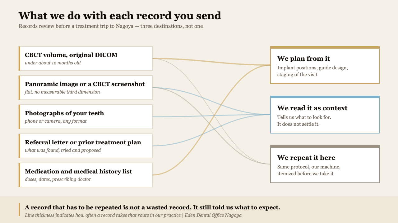
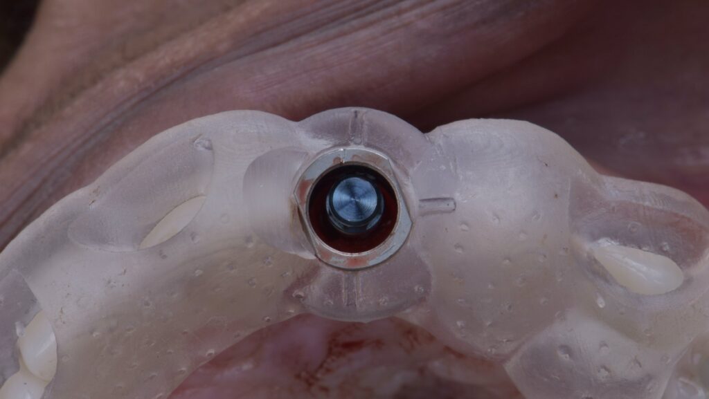
When a diagnostic cone beam volume arrives in its original form, we import it into our planning software, place the Straumann BLX or TLX positions virtually, and print a surgical guide in our in-house lab before your flight. That can turn a two-trip case into one. What records cannot do is fix the plan. We will give you a range and a visit structure, and a written estimate in English before anything begins — but the definitive plan follows the examination, not the email. A clinic that returns a complete plan and a firm price off one attachment is describing confidence it has not earned, as our note on what an implant quote should include sets out.
The five records worth sending, in order
Ranked by how much each changes what we can do.
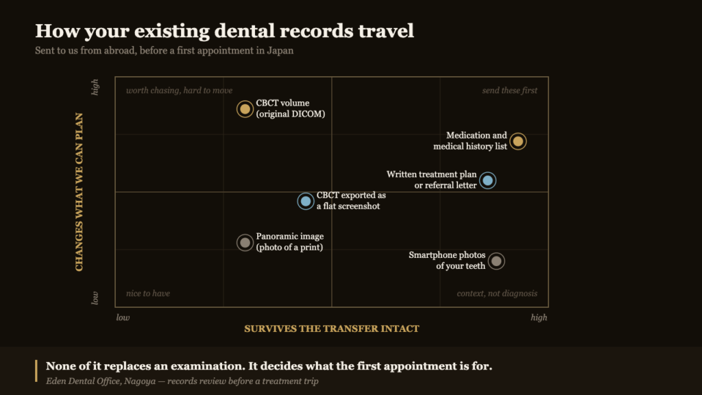
The cone beam volume, exported as DICOM. Ask for the original export — a folder of several hundred files, or a compressed archive, often 100 MB to over a gigabyte. Hard to move, and worth moving. Why a volume is a different instrument from a panoramic image is covered in our article on CBCT scanning before implant treatment.
Two-dimensional radiographs. Panoramic and periapical images, as files where possible. They date the problem, show previous root canal work and retained roots, and indicate whether bone loss is generalised or local.
Photographs of your own teeth. Four are enough: a relaxed smile, a full smile, and the upper and lower arches. No filters.
The referral letter or your previous treatment plan. What was found, what was tried, what was proposed, and the reasoning.
Your medication and medical history list. Generic drug names, doses, start dates. Anticoagulants, bisphosphonates and denosumab, diabetes control and smoking all change surgical sequencing, and this record travels perfectly in a plain email — see medications and implant treatment.
| Record | What it can settle before you fly | When it stops being usable |
|---|---|---|
| CBCT volume (DICOM) | Bone volume, nerve position, whether a guide can be printed in advance, how many surgical visits | Roughly 12 months, or immediately after any extraction, graft or new symptom at the site |
| Panoramic or periapical images | Existing restorations, retained roots, the broad pattern of bone loss, what to ask about | Anything treated or extracted since; a photograph of a print loses fine detail |
| Intraoral and smile photographs | Aesthetic starting point, smile line, gum display, how urgent the front teeth are | They date quickly with wear, staining and any new restoration |
| Referral letter or prior plan | What has already been tried, and why the case became difficult | Rarely expires; it is history, and history keeps |
| Medication and medical history | Surgical sequencing, bleeding management, whether a physician should be consulted first | Any change in prescription — send an update rather than a correction on the day |
The JPEG question, answered honestly
Patients apologise for sending a JPEG almost as often as they send one. The apology is mostly unnecessary. A 2024 scoping review in Brazilian Oral Research mapped eighteen studies published between 1996 and 2022 on whether image file format changes dental diagnosis; JPEG appeared in every study, TIFF in ten, and in most diagnostic tasks the format made no significant difference. More than seventy per cent of those studies carried a low risk of bias, and the authors concluded that any of these file formats can be used without impairing diagnostic accuracy.
Two limits on that finding are why your scan may still be repeated. Every study used intraoral images — twelve periapical, six bitewing — and none included DICOM. So compression rarely ruins a flat radiograph. What a flat picture cannot do is stand in for a volume, for a reason unrelated to file size: a panoramic image has variable magnification across the arch and no measurable third dimension. You cannot read bone width from it, or measure the distance from a planned implant to the inferior alveolar canal. That, almost always, is why a scan gets repeated — not a judgement on the image you sent.
What photographs can and cannot do
Photographs are the cheapest record to produce and the easiest to over-read. A 2024 systematic review of nineteen in-vivo studies in BMC Oral Health examined how accurately caries can be diagnosed from camera and smartphone photographs against clinical examination. Across those studies sensitivity ranged from 48 to 98.3 per cent and specificity from 83 to 100 per cent, with heterogeneity high enough that the authors declined to pool the results.
Read that the way a clinician does. High specificity means that when a photograph shows something, it usually is something. A sensitivity floor of 48 per cent means a clean set of photographs cannot tell you nothing is wrong. Useful for confirming, weak for excluding. Where they earn their place is aesthetics: smile line, gum display and the colour of the tooth beside the one being replaced, as our article on matching the shade of a single front tooth describes.
The record most often missing is written, not visual
The images arrive; the story does not. That pattern is not unique to international cases. A 2025 service evaluation at the Eastman Dental Institute in London reviewed 673 referrals from general practice and found that 160, or 23.8 per cent, were rejected at triage — and 148 of those rejections, 93 per cent, were for a lack of clinical information. Those were domestic referrals between clinicians sharing a language and a health system. The setting does not transfer; the mechanism does. What fails is rarely the imaging.
So ask your dentist, in writing, for the diagnosis as they see it, what has been attempted and when, the materials and implant systems already in your mouth, and what they would propose next. In most countries you can request a copy of your own records. If you are travelling because two clinicians have told you different things, our article on how a dental second opinion works in Japan covers what we do with a disagreement.
How to send them
A cone beam export will not fit in an email; we send a secure upload link and you drop the archive into it. Avoid messaging apps for the imaging itself, since most recompress it, and do not leave records on a public sharing link indefinitely. Once here, they are handled under Japan’s personal information protection legislation and live in the clinic record system — seen by Dr. Murai, your coordinator, and our in-house technician when a case is being designed.
What gets repeated here, and what that costs
Expect fresh intraoral photographs and a digital scan of your bite at the first appointment regardless of what you sent. They are the baseline every later stage is measured against. A cone beam scan is repeated when the volume is older than about a year, when the field of view misses the site, when artefact from existing metalwork obscures it, or when something has changed. Imaging is justified only when it will change what we do: if your scan answers the question, we use yours.
The published anchors on our fee page are the starting point — the examination and check-up package is ¥40,000, the whitening package ¥75,000, and annual membership runs from ¥88,000 to ¥288,000 by tier. Reviewing records you send before you travel is part of the consultation and is not billed separately. Any imaging we repeat appears as its own line on your written English estimate before it is taken, never afterwards. Where those records end up being read matters too: who holds the licence and what it permits is not something a clinic website usually makes clear. How Japanese self-pay fees compare internationally is set out in our comparison of dental costs across countries.
Frequently Asked Questions
Can you give me a treatment plan before I fly?
We can give a realistic range, a visit structure and an opinion on whether the trip is worth making. The definitive plan and firm figure follow the examination.
Is a photograph of my panoramic x-ray good enough?
For orientation, usually. For surgical planning, no: no flat image carries measurable bone width or nerve position.
How old can my CBCT scan be?
About twelve months, and it resets after any extraction, graft, infection or new symptom in the area. Planning from a volume that predates a change in the bone is the error we are avoiding.
Do my records need to be translated?
Radiographs and scans are language-neutral. For medication lists, send generic drug names with doses rather than brand names, which differ between countries.
Will I be charged twice if you repeat a scan?
You pay once, here, for what is taken here, quoted in writing beforehand. If your existing scan is adequate, we do not repeat it.
Records sent in advance reduce uncertainty; they do not remove it. A volume can look adequate on screen and prove unusable once we examine the site, and some cases need more visits than the images suggested. Surgical treatment carries risk in every setting — bleeding, infection, swelling, temporary numbness, sinus involvement where the sinus is close, and the possibility that a graft or implant fails to integrate. Timelines and costs here are typical and shift with the case. Individual results vary.
If you are considering treatment in Japan, send your records before you book anything. We will tell you what they settle and what they do not — see how we work with patients travelling from overseas, or message the clinic on WhatsApp in English. The conversation that follows is described in what a video consultation can honestly settle.
More on this topic: Trip Planning Guides — every article we have published in this cluster, grouped by stage.